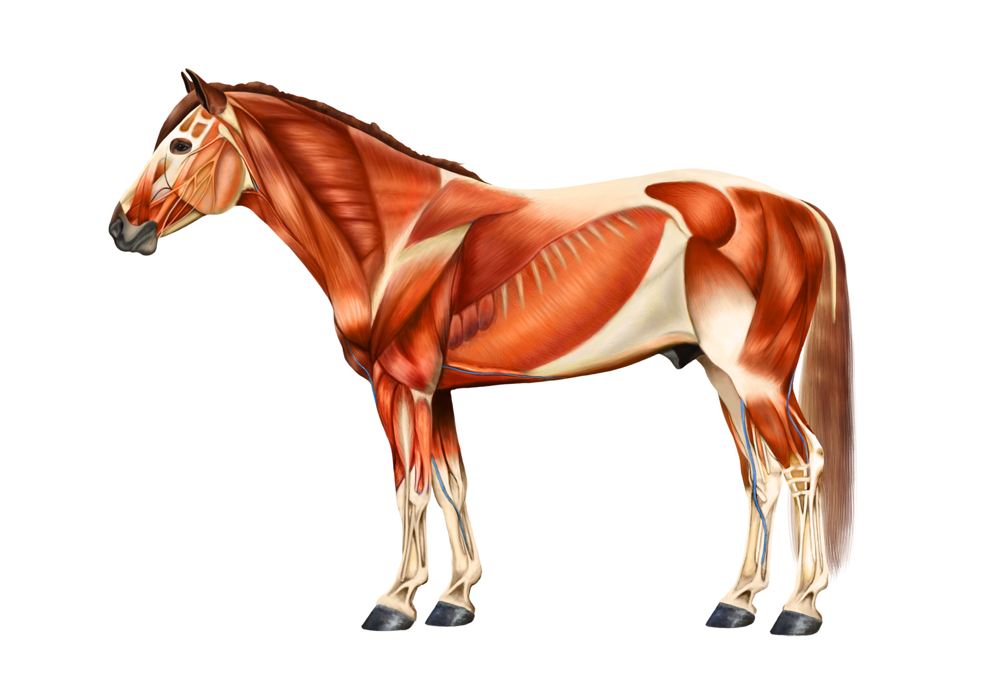
ArtStation Horse muscle anatomy
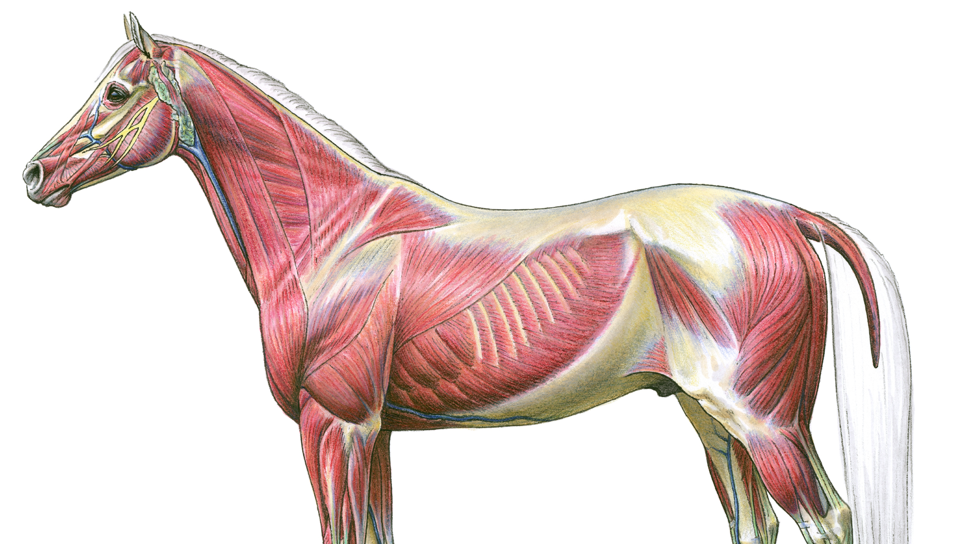
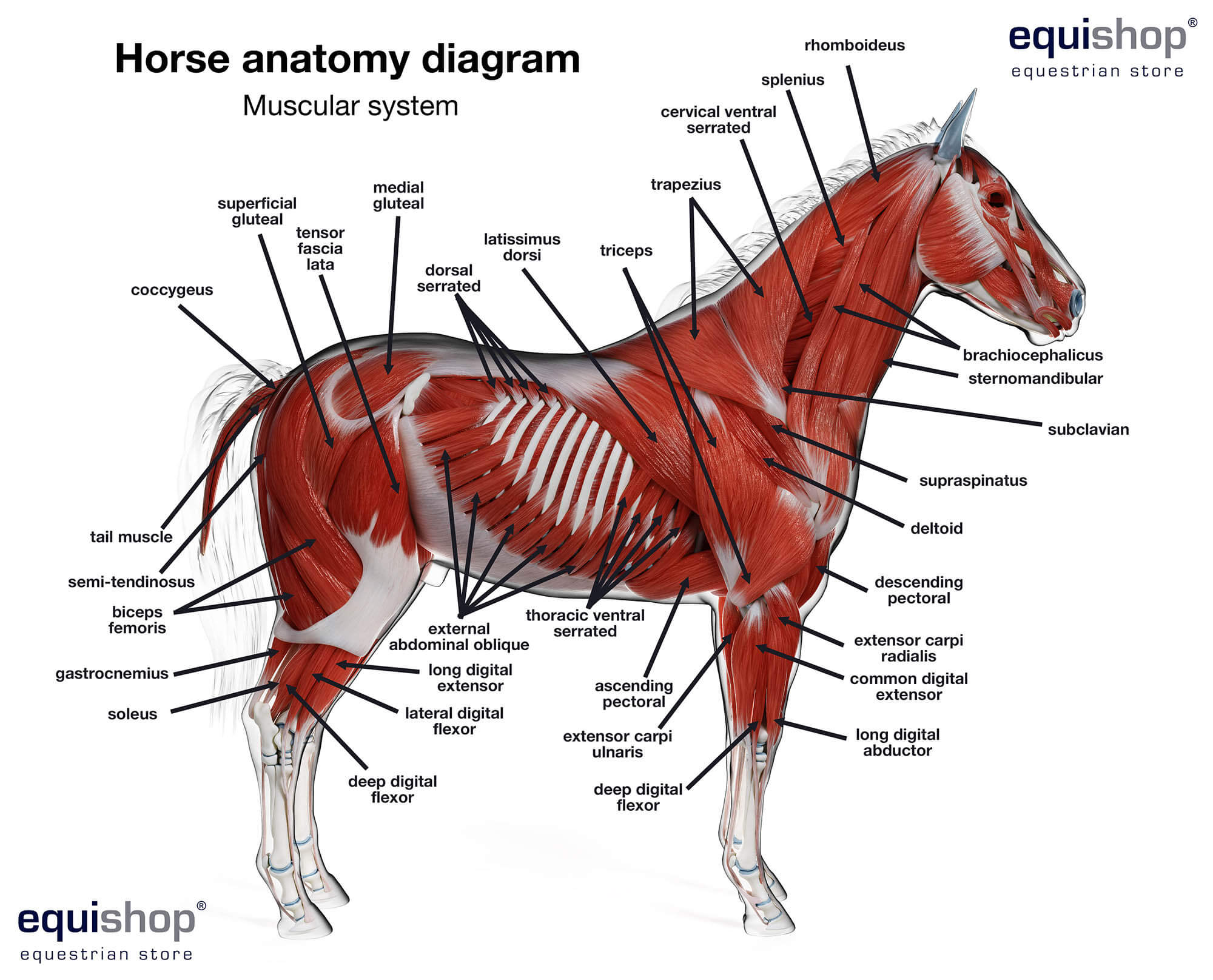
The longissimus dorsi, is the main muscle in the horse's back and underneath the saddle. However, it is not just local to this area. The longissimus dorsi starts at the 4th neck vertebra and and attached into the sacrum in the hind quarters. Branches of the longissimus dorsi also connect to the head and tail.
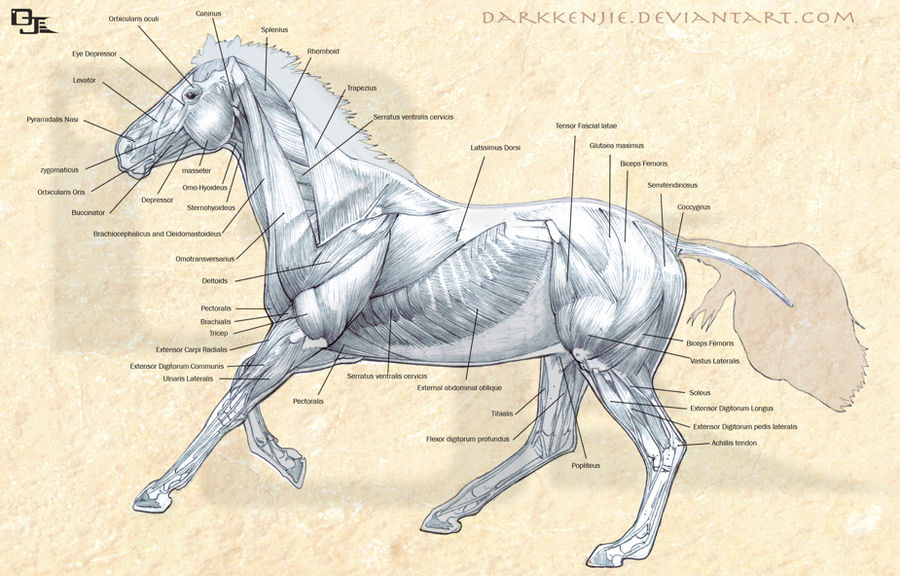
Horse Muscles Anatomy by DjWelch on DeviantArt
A muscle or muscles and its/their tendon (s) that operate together to cause flexion or extension of a joint are referred to respectively as a flexor unit and an extensor unit. Smooth: muscle which makes up automatic systems (digestive system, for example)

Horse Muscular Anatomy Poster
Wing of atlas and mastoid process of temporal bone. Elevates head and neck. Bends head and neck laterally. Stabilizes and extends vertebral column. Dorsal branch of local spinal nerve. M. Semispinalis capitis. Articular processes of C2/3-7 and transverse processes of T1-6/7. Occipital bone. Elevates head and neck.
Equine Muscles & Tendons
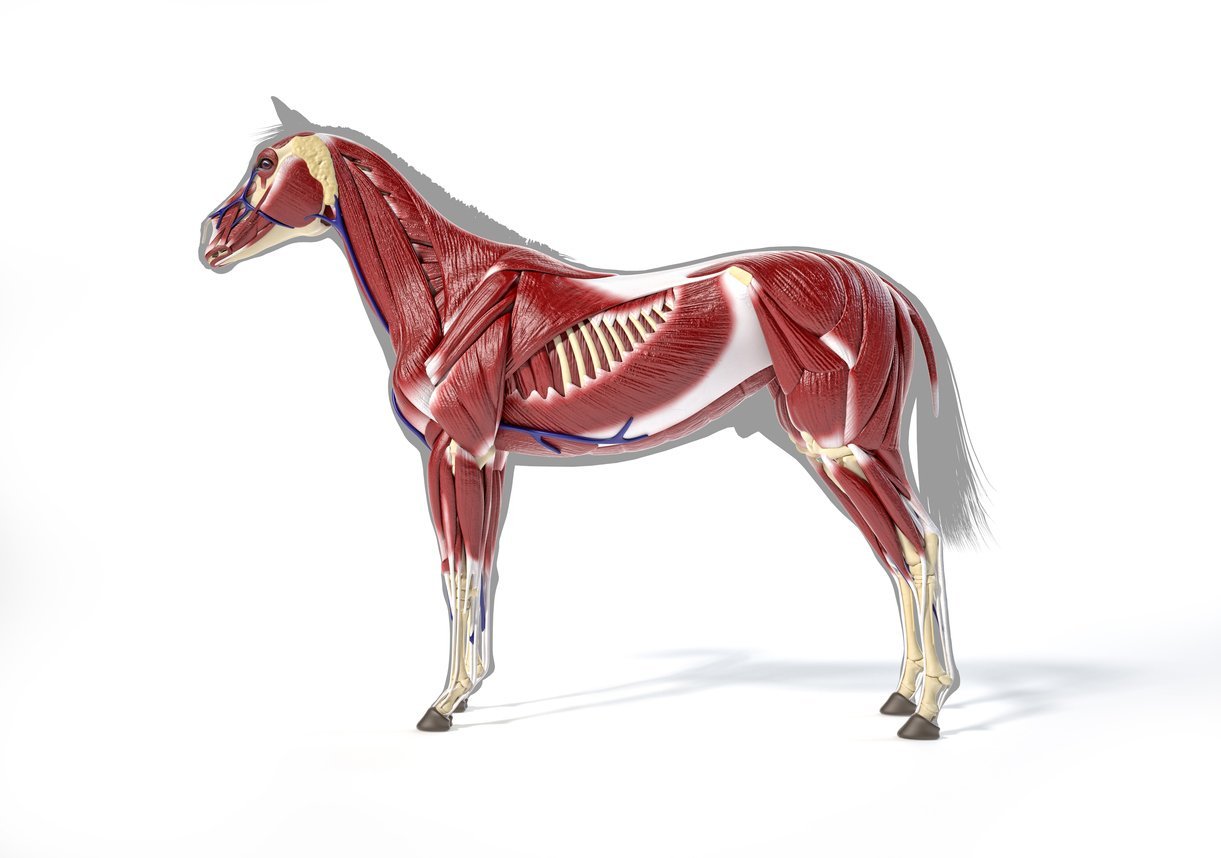
Horse Muscles. The muscular system of the horse is made up of skeletal muscle, smooth muscle and cardiac muscle.These are some of the main skeletal muscles in the horse and their function.. Brachiocephalicus, sternocephalicus and splenius help with the movement of the head and neck.Brachiocephalicus also extends the shoulder joint. Movements of the shoulder joint are facilitated by the.

Horse Anatomy the Muscles by COOKEcakes on DeviantArt
Dr Christin Finn, DVM, CVA details the underlying skeleton and muscle structure of a horse by using a live painted horse Find more information about your hor.

Image result for horse muscles diagram Horse anatomy, Dog anatomy, Horses
Anatomy of the Equine Skeleton. The horse skeleton consists of 200 different bones in the head, body, and legs. On the inside, every horse has the same horse parts, from the bone structure to the ligaments and horse muscles. But the size and look of the outer system can vary by equine race and gender. Horse Head
Horse Muscular System Poster Clinical Charts and Supplies
The horse's musculoskeletal system consists of the bones, cartilage, muscles, ligaments, and tendons. Their primary function is to support of the body, provide motion, and protect vital organs. There are 205 bones in the horse's skeleton.
Horse Muscular Anatomy Poster
The horse's body possesses approximately 700 muscles that control movement. Skeletal muscles, which attach to bones via tendons, contract or shorten in length in highly coordinated ways to.
Complete Guide on Horse Muscle [Domestic & Sport]
Croup and tail Horse skin and its products Summary While analyzing each part of the horse's body, we will also speak of the exterior - namely, the conformation, which depends on the horse's type and race. In this article, we will also speak about the horse's skeleton and muscles.
Anatomía del caballo diagramas de las partes del cuerpo del caballo Tienda Ecuestre
Horse Anatomy and Muscle Diagrams - stretchyourhorse 70% OFF! 25 VIDEO TUTORIAL COLLECTION Horse Anatomy and Muscle Diagrams This page contains color coded pictures of the horse's deep, superficial and hind end muscles. The pictures will help you "see" which muscles you are stretching!

animal muscles Cerca con Google Anatomie, Pferd, Reiten
Equine anatomy encompasses the gross and microscopic anatomy of horses, ponies and other equids, including donkeys, mules and zebras.

Superficial Front Limb & Neck Muscles, Front View Horse anatomy, Horse care, Horses
Horse Council . Title: Equine Anatomy Author: Essie Rogers Created Date: 12/28/2009 10:47:06 AM.

Equine Anatomy 3 Poster Collection Horse Organs Bone Muscles Chart
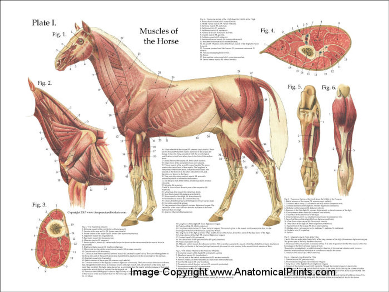
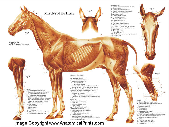
Microsoft Word - Muscles of the Horse and Their Purposes.doc Muscles of the Horse and Their Purposes http://www.asaddlery.com/info/anatomy.htm#Muscles Muscles of the forehand the jaw humerus forward, raises it in collection, swings the foreleg forward. rein contact stops free forward movement. Nuchal ligament Thoracic vertebrae Deltoid Poll Withers

Training Your Horse from the Anatomical Perspective. Part 2 Engagement
Large and only weight bearing component of crus (stifle/ knee) Large tibial tuberosity - patellar ligament Medial tibia is subcutaneous Cochlea is inclined craniolaterally. This causes the lower limb to move laterally on flexion Fibula is greatly reduced Distally incorporated into tibia Proximally tightly articulated with tibia It has a short shaft

THE ACTION OF MUSCLES
Jan 5, 2021 4 min read The Equine Muscular System The muscles of the body are responsible for creating movement whether it be via the skeletal muscle, smooth muscle, or cardiac muscle. Agonist muscles contract and are the primary mover.

Equine deep musculature anatomy chart Horse anatomy, Equine veterinary, Muscle anatomy
#1. Osteology - horse bones anatomy #2. Myology of horse - special properties of horse muscles #3. Digestive system - exceptional anatomical features of the digestive tract of a horse #4.